Quantifying the uncertainty of Electric Properties Tomography in medical imaging
Challenge
Magnetic Resonance Imaging (MRI) is a diagnostic, mainly qualitative, technique used to visualise the soft tissues of a patient. The application of a strong magnetic field causes the protons in a patient’s hydrogen atoms to align with it. Radiofrequency pulses are then used to force the protons to move out of and back into alignment with the field, generating signals characteristic of the tissues in which the hydrogen atoms reside. These signals are interpreted by software to provide an MRI image. Contrasting agents, often metals such as gadolinium, are used to enhance signals. Although generally safe, evidence suggests these agents can build up in the body and, on rare occasions, cause adverse reactions in some patients.
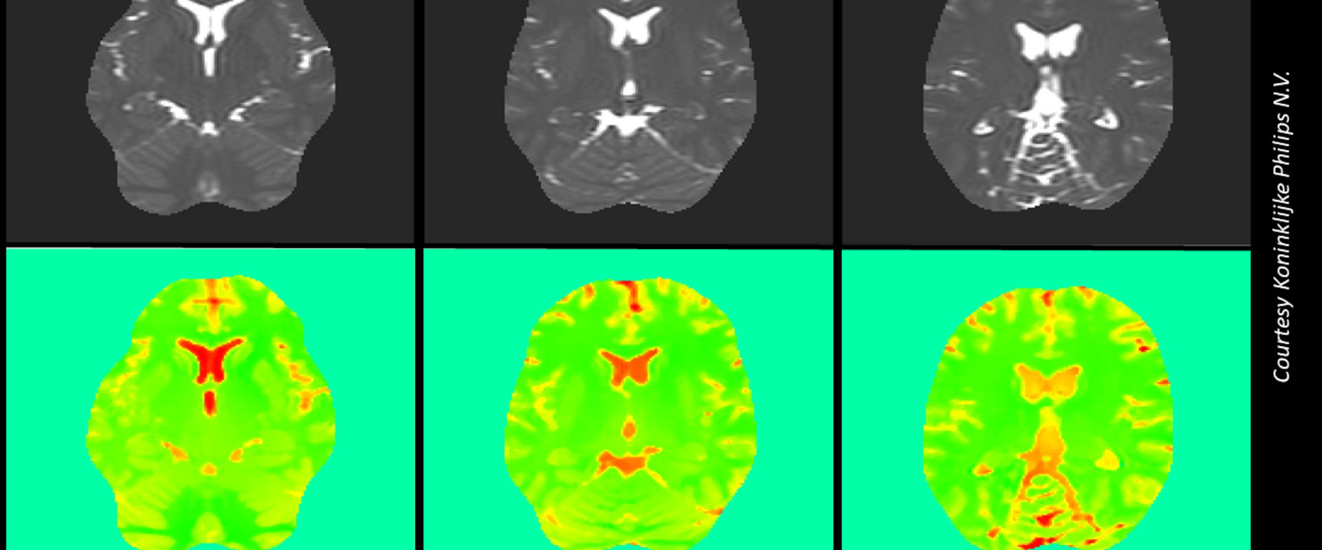
Electric Properties Tomography (EPT) is a MR-based method that does not require contrasting agents. Different body tissues have different electrical conductivities that interact with the scanners’ radiofrequency magnetic field, causing fluctuations that can be interpreted to provide quantitative data.
However, various factors can affect measurements – such as the instrument, operator or environment – quantified by measurement uncertainty (MU), indicating the quality of the measured value. No MU assessment had ever been applied to EPT during its development, meaning that comparing data from one scan to another was not metrologically valid.
Solution
During the EMUE project, the first MU assessment for the commonly used Helmholtz-EPT technique was performed. To avoid the uncertainty arising from different tissue types, a homogenous phantom was constructed containing salt water to mimic the average electrical conductivity of the human body. Twenty-five scans were performed with the phantom stationary in a Philips 3 T Ingenia TX MRI instrument to provide repeatability measurements without the uncertainty due to placement. The analysis allowed accounting for many sources of uncertainty, such as the measurement fluctuations of the MR signal and imaging artefacts.
Measurement uncertainty was propagated through the mathematical model implemented in EPTlib, open-source software developed in the context of QUIERO, a project developing metrology for quantitative MRI. A major factor affecting the uncertainty propagation was the choice of kernel used, which defines the mathematical ‘smoothing’ applied to the voxels (the 3D representation of the pixels in scan images) during image processing.
The study revealed that the repeatability uncertainty was very small - indicating good repeatability for this technique. However, spatial fluctuations were observed in the obtained conductivity images, indicating the need of a more extended uncertainty assessment. To this end, a further set of scans of the phantom at different positions were performed in the QUIERO project to assess the MU associated with reproducibility. The resulting reproducibility uncertainty was in good agreement with the observed spatial fluctuations of each single scan.
Impact
Philips, a world leading provider of healthcare solutions and specialists in medical scanning, generated the phantom and performed the MRI scans in the EMUE and QUIERO projects. The company’s involvement in these was motivated by their core belief that through innovation “there’s always a way to make life better”. Philips had previously participated in EPT research due to the ability of its instruments to discriminate between benign and malignant tissue in cancers such as breast and brain, providing the possibility of minimising unnecessary surgery or earlier intervention in pre-metastatic cancer.
Although only the first steps have been taken to provide a full, rigorous, measurement uncertainty in EPT applications, these concepts will be extended using the approach developed in the EMPIR projects. Knowledge of the different electric properties of in vivo tissues for EPT will allow pathologies to be monitored over time without the use of contrast agents and provide greater trust in the use of scans and scanning instruments.
- Category
- EMPIR,
- Health,
- Standardisation,
- EMN Mathematics and Statistics,
