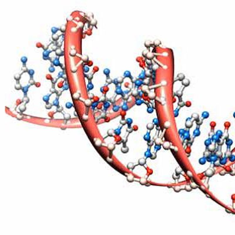
Simulating the effectiveness of radiotherapy at the nanometre scale
Challenge
One in two people will develop cancer in their lifetime. Radiotherapy, using external sources of radiation, is a common treatment approach. This includes photon sources, such as x-rays and γ-rays, and particles, such as electrons, protons, and carbon ions. Radiation doses must be carefully calculated – too high a level and healthy tissue can be affected, too low and the cancerous cells may survive.
When radiation beams hit a cell energy is deposited, mainly through the production of secondary electrons which interact with the molecules in the medium. These interactions form nanometre scale patterns or ‘particle track’ structures which correlate with DNA damage. Different radiation types have different particle track structures and damage the DNA to a different extent, resulting in different biological effectiveness. Chemical changes ensue, including the formation of free radicals which also attack the DNA. This damage activates cellular repair mechanisms and influences cell survival.
Patient treatment plans aim to minimise the risk of adverse effects but often consider only the average radiation dose at volumes of millimetre dimensions, orders of magnitude higher than the nanometre-scale diameter of the DNA – considered to be the primary radiation target.
For particle beams different approaches are applied to account for the enhanced biological effectiveness. As a result, different centres employing the same treatment on the same condition can use up to a 20% different radiation dose.
Solution
During the EMRP BioQuaRT project, the French Institute for Radiological Protection and Nuclear Safety (IRSN) developed a DNA damage simulation chain, based on Geant4-DNA, an extension of the Geant4 Monte Carlo toolkit for track structure calculations and biological damage simulation.
An essential ingredient of these calculations are the ‘cross-sections’ for the interaction of radiation with the molecules in the medium. Prior to the BioQuaRT project, only water cross-sections were used in the Geant4 DNA code. In the project, cross-sections for the interaction of radiation beams with DNA constituents were measured.
Results were implemented into a new Geant4-DNA design and validated using the nanometric scale Monte Carlo code Ptra. A new geometrical DNA mode was implemented to include both direct energy depositions on DNA and subsequent free radicals on DNA damage. The long computational time required for the chemical stage limited models to a 30 nm diameter chromatin fibre, thus scaling procedures were used to extrapolate to a cell nucleus.
To validate the results in terms of early DNA damage (double strand breaks and chromosomal aberrations) experimental data was obtained from radiobiological experiments on endothelium cells.
The improved Geant4 simulation chain was able model the physical and chemical changes of ionising beams down to the diameter of biological molecules inside cells.
Impact
At the end of the project in 2015, IRSN, as member of the Geant4- DNA collaboration, further developed the code. In 2017 this was extended to incorporate modelling of the physical, physicochemical, and chemical stages of early radiation damage at the scale of an entire human genome. This was publicly released to the Geant4 community. In 2021, a new method termed IRT-sync, developed in the frame of the Geant4-DNA collaboration, was added to allow a faster simulation of the chemical stage, later extended to include DNA repair models.
The model can now compare the risks of one beam type to another and its predictive capabilities can replace complex and costly experiments.
IRNS acknowledge the help of the EMRP project in developing the code, especially the access to knowledge and laboratories of project partners such as PTB, the National Metrology Institute of Germany, who precisely measured the cross-section data of DNA constituents and gave access to its microbeam facility for radiobiological experiments.
The code will be extended in the future, validating it against clinical proton or carbon ion therapy treatments and extending it to cover new therapy types such as flash therapy. The new code will help radiotherapy plans, which consider both physical dose and the predicted treatment outcome. This will enable results from different radiotherapy centres in Europe to be more easily compared, reduce the need for expensive radiobiological cell and animal experiments, and provide better treatment for patients.
- Category
- EMRP,
- SI Broader Scope / Integrated European Metrology,
